In Dermatology, a biopsy is a commonly performed procedure. Because we are a medical field that specializes in the detection and treatment of skin cancer, a biopsy is our way of making a proper diagnosis. Biopsies are in no way unique to dermatology, and, in fact, you may have had one before from other body areas (ex: a breast biopsy for a new lump, or a biopsy of a suspicious polyp or growth during a colonoscopy, etc…)
A biopsy entails sampling a small piece of tissue to send for pathologic testing. This article details the steps that occur from start to finish.
- Usually a skin cancer is detected one of two ways: the patient comes in to REN Dermatology specifically reporting a new, growing, or changing skin spot (what we call a “lesion”), or a patient comes in for a routine yearly skin cancer screening visit and during the exam, we notice a lesion that appears concerning.
- Once we’ve identified the suspicious lesion, we recommend a biopsy. In general, skin biopsies are very quick, relatively painless and require minimal downtime. First, we clean the area with a swipe of alcohol. We numb the area with a small injection of an anesthetic (typically Lidocaine mixed with Epinephrine*). There is a slight stinging sensation with the Lidocaine injection, but this is momentary and lasts just a few seconds. The Lidocaine is mixed with Epinephrine which helps to minimize bleeding. I should also mention that we inject the Lidocaine with a 30 gauge needle (very tiny!)
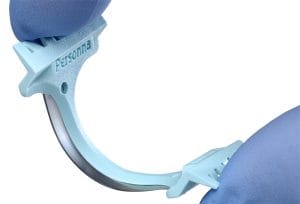
- Now that you’re properly anesthetized, the actual biopsy procedure should not hurt at all and will actually take less than a minute. In most cases, we will do what’s called a Shave biopsy. We use a flexible blade to shave off the spot of concern.

Shave Biopsy Tool: Dermablade
- There is typically very little bleeding, but we use a medicated solution that is able to stop any bleeding immediately. Once all bleeding has stopped and the resulting wound is clean and cry, we apply plain petrolatum jelly and a small bandage.
- Occasionally we will do a Punch biopsy. This utilizes a tool that cores out a small piece (usually 4mm in diameter) of tissue, after which we place two stitches to close up the area. A punch biopsy is often used to help evaluate rashes or lesions concerning for Melanoma.
Read on to learn more about what happens AFTER the biopsy
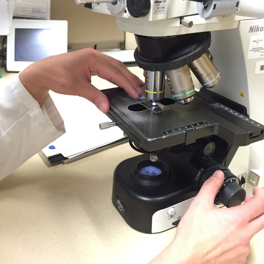
Your specimen is placed in a sterile bottle containing formalin. At the end of the day, all the specimens are picked up and brought to a laboratory for processing. The processing happens overnight and allows your biopsy sample to be sliced into very thin layers, stained with special coloring stains, and placed on a microscope slide. This slide then goes to the pathologist, a board-certified doctor who is an expert in reading and interpreting pathology slides.
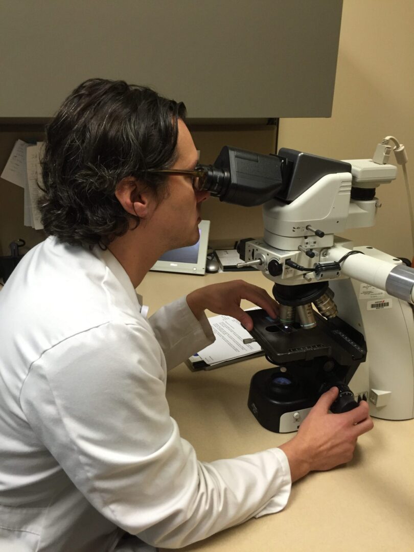
Dr. Jeffrey Zwerner, Vanderbilt Dermatopathologist
At REN Dermatology, we send all our specimens to Vanderbilt, specifically to the dermatopathologists. These doctors are additionally fellowship-trained to specialize in reading biopsies of skin. We feel this provides the highest-level of interpretation of your biopsy. In less than a week, we will have the results from your biopsy. The pathology report (or “biopsy result”) helps guide us in choosing the best treatment for your condition or diagnosis.
As the patient, you will likely never come into contact with the dermatopathologists, but they are essential in helping us take great care of you. We work collaboratively with them to interpret your results and often rely on them to make an accurate final diagnosis.
Click here to read about skin cancer.
By: Dr. Jennifer Lee